The muscles have been studied prior so its essential you have a base understanding of the muscoskeletal system before beginning.
Enjoy...
Enjoy...
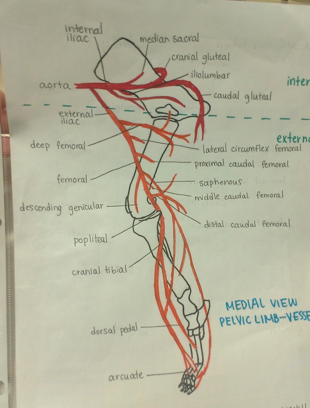
The first image is the vessels of the pelvic limb. We follow the external iliac artery (branch of the aorta) out of the vascular lacuna where it becomes the femoral artery. The femoral artery has seven (7) branches. After the distal causal femoral artery the femoral a. turns into the politeal a. and as it continues down it becomes cranial tibial a. and finishes as dorsal pedal and arcuate a.
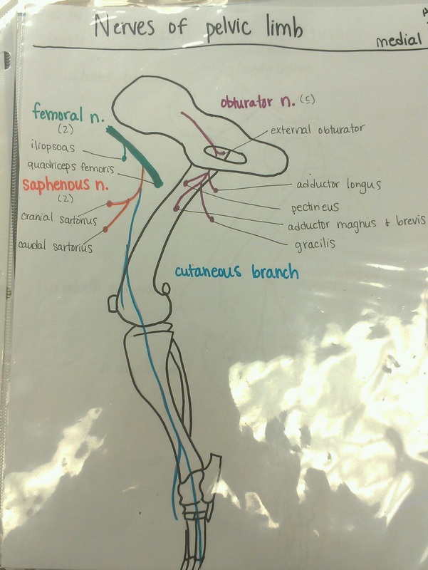
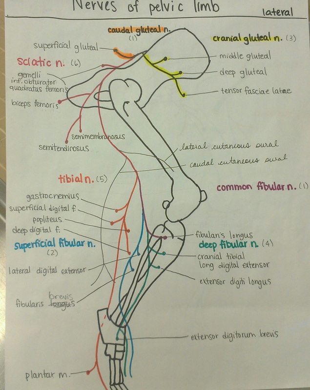
These two pictures show the medial and lateral nerves of the pelvic limb. Nothing too tricky here.
Note: Common fibular n. branches into two the superficial fibular n. and deep fibular n.
Note: Common fibular n. branches into two the superficial fibular n. and deep fibular n.
 RSS Feed
